Create a free profile to get unlimited access to exclusive videos, sweepstakes, and more!
Cerebral cartography! Building the Google Earth of brain maps
Hey, Google, give me directions to the hippocampus.

In the 2015 film Self/Less, Damian Hale, played by Sir Ben Kingsley undergoes a cutting-edge procedure to transfer his consciousness into another body. The specifics of the operation are left intentionally obscured, instead treating the audience to some fancy looking technologies which capture all the bits of Hale’s brain and move them into his new shell.
There are, of course, important ethical questions surrounding whether or not we should embark upon that sort of technology if and when it becomes available to us. Admittedly, we’re nowhere near having a robust enough understanding of the brain’s machinations to make a workable copy of it, either inside of another body or a computer, but we might be about to get a bit closer.
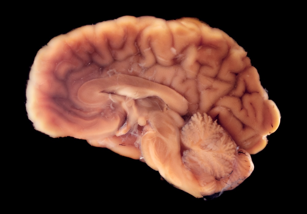
The brain consists of billions of individual cells, all working together in complex ways to manage your body’s maintenance and create your conscious self. Before we can even dream of duplicating the brain’s inner workings, we’ll need to have much better maps showing us how things are laid out, not just in one brain, but across the vast diversity present inside the skulls of human beings.
Recently, the National Institutes of Health awarded a $9.1 million grant to researchers at Columbia and the Icahn School of Medicine, to develop better and faster brain mapping techniques.
Today, mapping a complete human brain is a painstaking process which can take years to complete. The technique under development by Elizabeth Hillman from Columbia’s Zuckerman Institute, Zhuhao Wu from Mount Sinai’s Laboratory of Neural Systems, Structures, and Genetics, and colleagues, is called HOLiS — which stands for Human Brain Optimized Light Sheet — could cut the mapping down to a matter of days or weeks.
“There’s been super growth in being able to do this on the scale of a whole mouse brain, but everything goes horribly wrong when it’s a human brain because it’s ginormous. You could just say let’s take the human brain and chip it up into little cubes the size of a mouse brain and do it that way, but any time you’re cutting you get edges you can’t deal with and lost tissue,” Hillman told SYFY WIRE.
Current techniques require cutting the brain into incredibly thin slices and slowly imaging them. You then have to put all of that data together. The process can take years, and, in the meantime, the tissue is changing or degrading. HOLiS will allow researchers to slice the brain into comparatively large slabs roughly 5 millimeters thick. The whole brain could be broken down into only 30 cassettes, instead of hundreds or thousands.
“Even a mouse brain was challenging five or ten years ago,” Wu told SYFY WIRE. “If you wanted to see everything in a mouse brain, you’re talking about thousands of sections. We’re really leveraging recent technologies to get to this state-of-the-art approach.”
Once the brain is cut into slices, it is rendered totally transparent, allowing the microscope to gather data across the slice. Researchers will also use an array of staining techniques to tag particular structures, making them more readily apparent. From there, the microscope will go to work, gathering images at speeds not yet seen in brain imaging.
“In the past, people would take an image and move, then take an image and move. When you budget the millions of little moves, that adds up. Instead, we move as we image, so there’s almost no budget in our system for movement,” Hillman said.
The one-week imaging timeframe is based on a number of assumptions and likely won’t happen right out of the gate. Hillman said that the first brain will probably take longer as they establish the protocol, but it should speed up as the process finds its feet. That timeframe is also based on imaging one tissue cassette at a time and one brain at a time.
“The budget we calculated was assuming one system, so each slab would be done one after the other. We could be doing them at the same time on a bunch of systems,” Hillman said.
Having high-quality reference maps of human brains won’t be used for high-flying science fiction consciousness transfer — at least not yet. Instead, they will serve as a foundation of knowledge for understanding how brains work across different age ranges and physiological conditions.
“We don’t understand how one brain differs from another. You could take a chunk of someone’s brain out who has epilepsy and image a bunch of cells, but we don’ know enough even baseline to understand what it would mean,” Hillman said.
That’s ultimately what the project is hoping to address. “There’s so much diversity. We want to image hundreds or thousands of human brains. We’re carefully designing the first cohort of brains we image to cover enough diversity from different ages, different diseases, and controls. That’s the first step we need to do to understand how much diversity is there,” Wu said.
Having a collection of reference brains, all representing structural maps at different stages and under different conditions, would give doctors and scientists something to bounce real-world scenarios up against. They could image a patient and compare it to the reference to get an idea of what might be going on inside a particular person’s brain. The reference maps could also serve as an educational resource to a wider audience, allowing people to virtually navigate the brain’s various structures and see how they work together.
“I could share all the satellite images of the surface of the Earth with you and what would you have? Just tons of images. What you need to have is something that tells you what’s a building, what’s a cow, and what’s a road. What we’re trying to understand is what people will use this information for and give them that readout,” Hillman said.
The information present in even a single brain is staggering. The team estimates that each brain will produce roughly two petabytes of data, all of which needs to be put together into a format which can be used by doctors, researchers, or curious people on the internet. Once the system is up and running, researchers aim to map reference brains with readily visible tags, such that the information is machine-readable.
If all goes according to plan, you’ll soon be able to explore the human brain at a cellular level. It isn’t body snatching immortality, but it’s pretty cool, and given the way Self/Less played out, it’s probably for the best.


