Create a free profile to get unlimited access to exclusive videos, sweepstakes, and more!
Watch out, Spongebob, because worms that keep growing new arms live inside sponges

Worms take many peculiar forms, from oversized lipsticks to brightly colored Christmas trees, but this might be the most bizarre one yet.
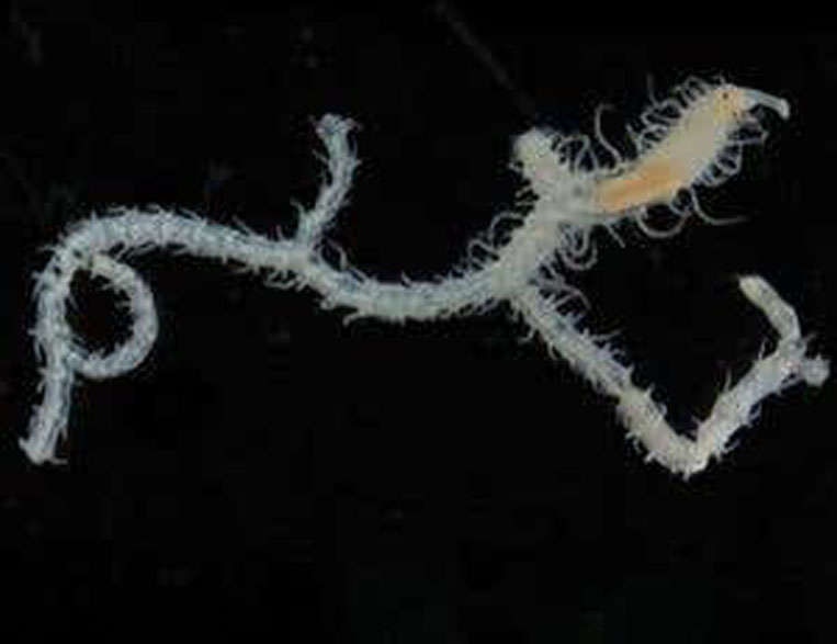
Spongebob would probably not be too thrilled to find out a worm was squirming through his entire body—though he probably would use it as an excuse to take a day off from Krabby Patty. Sponges are labyrinthine on the inside, and the worm Ramisyllis multicaudata or branching syllid worm may have only one head, but this annelid (segmented worm) is able to navigate by bifurcating, or splitting a section of its body in half for an instant arm whenever it needs one. It also reproduces by releasing stolons that have their own brains and eyes.
Officially discovered in 2006 by Chris Glasby, its insides have finally been made sense of by Glasby, Guillermo Ponz-Segrelles and an international research team who recently published a study in Journal of Morphology and proved that the weird only get weirder once you dissect them.
“The evolution of the bifurcating body of Ramisyllis multicaudata is related to its symbiotic relation with these sponges,” Ponz-Segrelles told SYFY WIRE. “Right now, our hypothesis is that the sponges are not so much related to the appearance of this body shape in the first place, but with the fact that the animals can survive despite their strange anatomy.”
Glasby is the senior curator of annelids (segmented worms) at the Museum and Art Gallery of Northern Australia. He had been assisting the museum's bioprospecting project that was seeking samples from marine animals that could possibly be used for future medications. Many of these samples, which the museum kept parts of before sending them off, came from invertebrates like sponges. When he noticed wormlike things emerging from the pores of a sponge, he had to take a closer look.
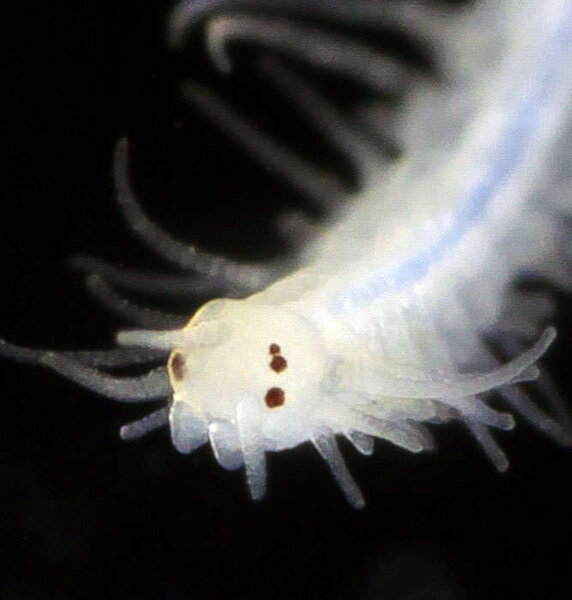
“I found that rather than many worms, the tails belonged to one, enormously branched worm,” Glasby told SYFY WIRE. “It took several days to find the head, through careful dissection of the sponge. Then I realized we had found only the second ever example of a branching annelid worm.”
This first dissection ended up in the worm’s body being broken, because which of its many arms led to the head was unknown, but the head was finally found near the base of the sponge. Glasby developed a technique for dissecting sponges so specimens of R. multicaudata could be extracted from any sponge they were found in. This time, he and his team have gone even deeper. Advanced microscopy and scanning techniques went where no scientist ever had before. X-ray-based micro-CT scans showed how the worms positioned themselves inside the sponges, something that could not otherwise be answered without ruining the worm, since you can’t exactly see through a sponge. The scans created thousands of virtual slices of sponge that revealed the worm’s whereabouts.
“The great thing about this discovery was the potential to research its strange body form and biology,” Glasby said.
Even that hi-res technique still wasn’t at a high enough resolution to see inside the worm and figure out where its organs were. The team used histology, which looks into microscopic structures within tissues and involves staining thin pieces of the specimen and photographing them so each organ can be detected. 3D reconstructions of the organs could then be created. They also used immunohistochemistry, staining the specimen with fluorescent antibodies that attach to molecules and are seen by a laser scanning microscope. This makes 3D reconstructions of the nervous system and others possible.
“Different anatomical techniques allow us to investigate different aspects of the animals with varying degrees of detail,” Ponz-Segrelles said.
Combining them gave us a complete perspective of how these tiny animals are structured on the inside at a remarkably high resolution in which we can sometimes detect even individual cells. The level of understanding that these combined methods offer could never be achieved with a single technique.”
Studying the branching syllid worm up close gave the researchers insight into its nervous system, which actually has its epicenter in the digestive system and branches out from there, as well as why something about the muscles of the worm looked strange wherever it branched off. Reconstructing the insides of the worm in 3D showed a previously unknown structure now known as a muscle bridge. Whenever a new branch grows from the side of the worm, the muscles in that side of its body stay where they are from these structures. Ponz-Segrelles and the rest of the team are now focused on finding out how exactly the intestines and circulatory system function in a worm that lives in a maze and is a maze in itself.
“Now that we have described the anatomy, we have all sorts of questions related to the functioning of these animals,” he said. But why do these creatures reproduce in such an alien way? Survival explains why the worm never leaves its host sponge and instead sends up stolons that can see and sense potential predators to avoid. If they are snapped up by something, the worm will still be alive to produce more stolons later on.
From the way they split themselves to how they propagate their genes, these worms are much more fascinating than anything that lives in a pineapple under the sea.